Fundus Photography
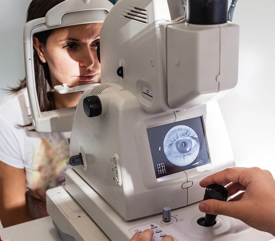
A fundus photograph is a specialized form of medical imaging. Using a customized camera with high-powered lenses that are mounted to a microscope, photographs are taken of the back of the eye by focusing light through the cornea, pupil and lens.
Fundus photographs are used to identify or monitor a wide variety of ophthalmic conditions. To begin the process, the pupil is dilated with eye drops. The patient will be asked to stare at a fixed device, keeping the eyes focused and still. There will be a series of flashes of light. The process usually takes no more than 10 minutes.
Some of the ophthalmic conditions fundus photography is used for include:
- Glaucoma
- Diabetic retinopathy
- Macular edema
- Microaneurysm
- Optic nerve
- Fundus photography has also been used to interpret the results of a fluorescein angiogram.
Contact us
Contact us today to learn more about Fundus Photography.
The doctors at Cincinnati Eye Institute have either authored or reviewed the content on this site.