
Retinal Vein Occlusion
A retinal vein occlusion is also known as venous stasis retinopathy or hemorrhagic retinopathy. It typically occurs in men and women over the age of 50.
What is a Retinal Vein Occlusion?
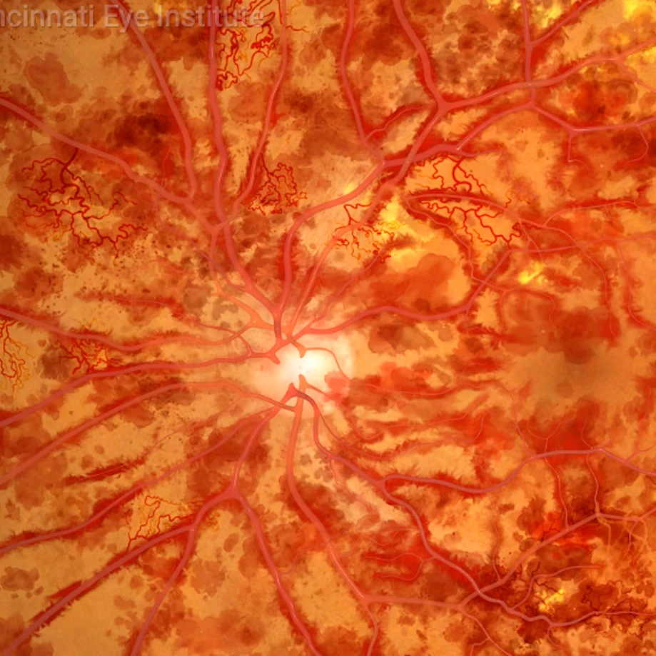
The retina is a thin sheet of nerve tissue located in the back of the eye where light rays are focused and transmitted to the brain. Tiny blood vessels supply the retina with oxygen and other nutrients. Arteries deliver blood and the retinal veins carry it. Sometimes one of these arteries hardens or swells and presses on a nearby vein. The vein can then become blocked, or occluded, making it difficult for blood to leave the eye. This condition is called a retinal vein occlusion, or RVO.
The blocked circulation caused by a retinal vein occlusion can lead to:
- Swelling
- Bleeding
- Growth of abnormal blood vessels
- Partial or total vision loss
Retinal vein occlusions are the second most common cause of blood vessel-related vision loss, the first being diabetic retinopathy. A retinal vein occlusion typically occurs in men and women over the age of 50, particularly those in their 60s and 70s.
Types of Retinal Vein Occlusions
Branch Retinal Vein Occlusion (BRVO) – A branch retinal vein occlusion is the blockage of a vein in the inner portion of the eye, where retinal veins “branch out” to smaller veins.
Central Retinal Vein Occlusion (CRVO) – A central retinal vein occlusion is a blockage that occurs in the central or main retinal vein located at the back of the eye.
Risk Factors for a Retinal Vein Occlusion
Risk factors for a retinal vein occlusion include:
- High blood pressure
- High cholesterol
- Diabetes
- Smoking
- Glaucoma
- Vitreous hemorrhage
- Macular edema
- Inflammatory conditions
Symptoms of a Retinal Vein Occlusion
Symptoms of a retinal vein occlusion include a sudden loss of vision or blurring of vision in all or a part of the eye.
Diagnosis of a Retinal Vein Occlusion
A retinal vein occlusion is detected during a retinal exam of the eye. After a thorough medical examination of the eye, the following diagnostic tests may be conducted to confirm the diagnosis of a retinal vein occlusion:
- Fluorescein angiogram
- Testing of intraocular pressure
- Pupil reflex response
- Slit-lamp examination
- Visual field testing
- Visual acuity
- Retinal photography
- Blood tests
The initial bleeding can prevent the ophthalmologist from seeing any other symptoms for three to six months or longer. The patient is monitored during this time until the blood clears.

Treatment of a Retinal Vein Occlusion
Treatment of a retinal vein occlusion depends on the severity and location of the blockage. Most patients vision will be restored but their vision is rarely is the same as it was before the occlusion.
There is no cure for a retinal vein occlusion. Emphasis is placed on prevention of the condition by treating the symptoms and preventing further vision loss. A retinal vein occlusion is an indication of vascular disease. It is critical to reduce the risks of vascular disease by adhering to the following guidelines:
- High blood pressure
- High cholesterol
- Diabetes
- Stop smoking
- Eat a diet low in fat
- Maintaining weight
- Exercising regularly
Complications of a Retinal Vein Occlusion
Complications of a retinal vein occlusion occur and require treatment that may include:
- Focal laser treatment for macular edema
- Intraocular injections of an anti-vascular endothelial growth factor
- Laser treatment to prevent the growth of blood vessels that can cause glaucoma
Meet Our Doctors


Schedule an Appointment
Contact us to schedule an appointment.
The doctors at Cincinnati Eye Institute have either authored or reviewed the content on this site.